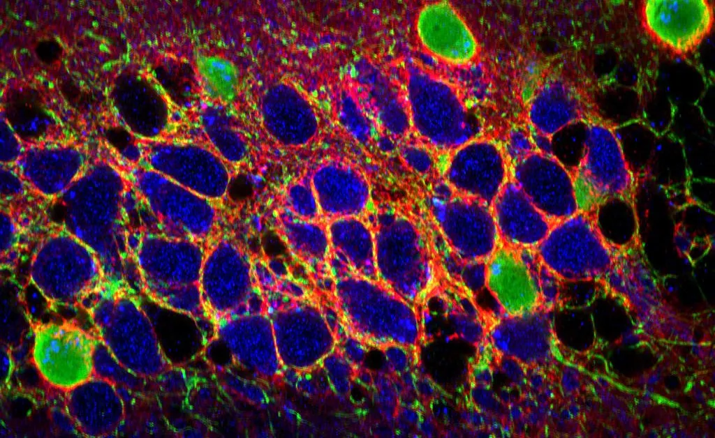
The perineuronal net extracellular matrix of the hippocampal CA2 subfield
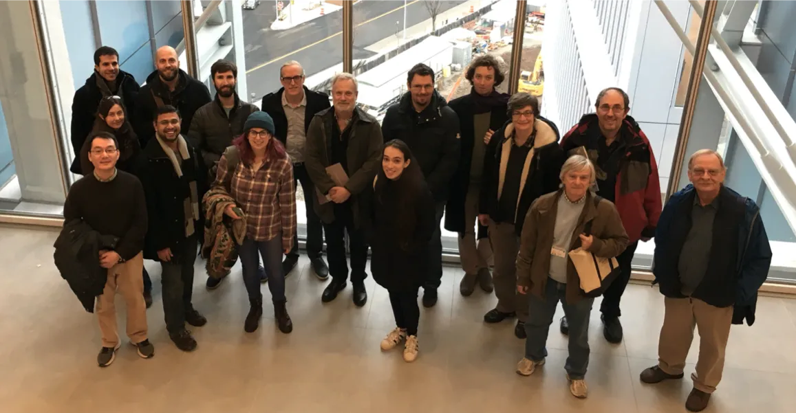
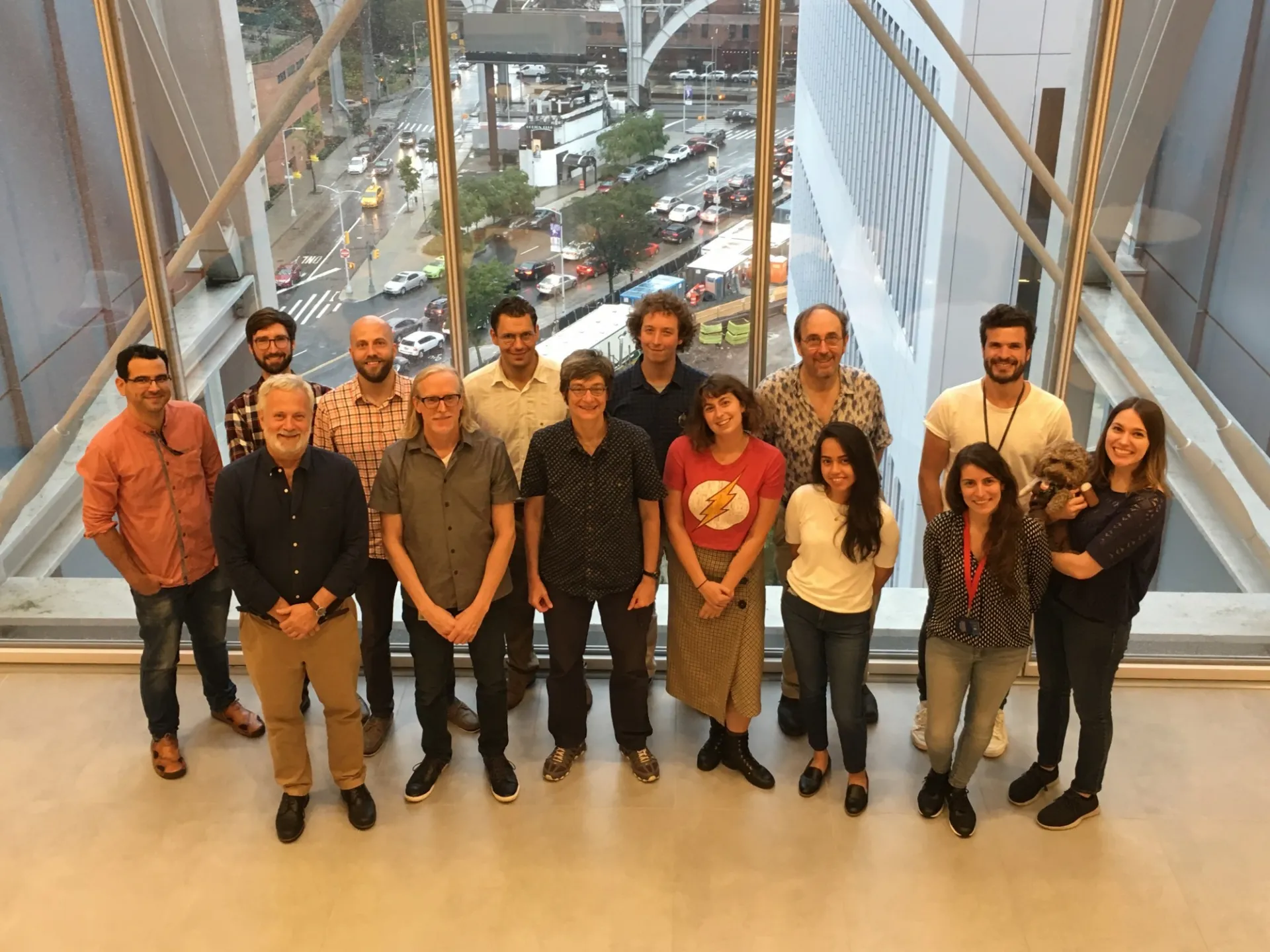
lab members 2019
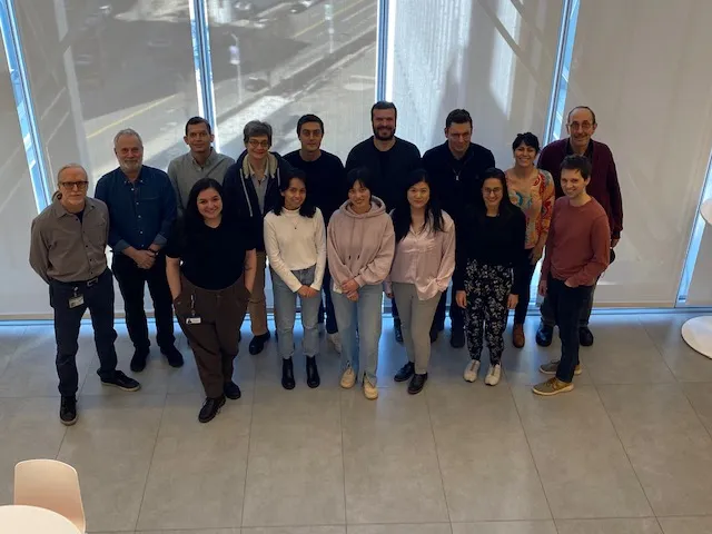
one of the Zuckerman Institute weekly happy hours
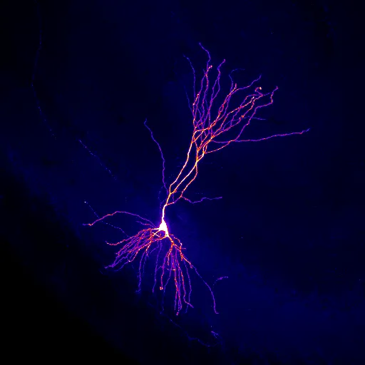
single biocytin-filled CA2 pyramidal neuron
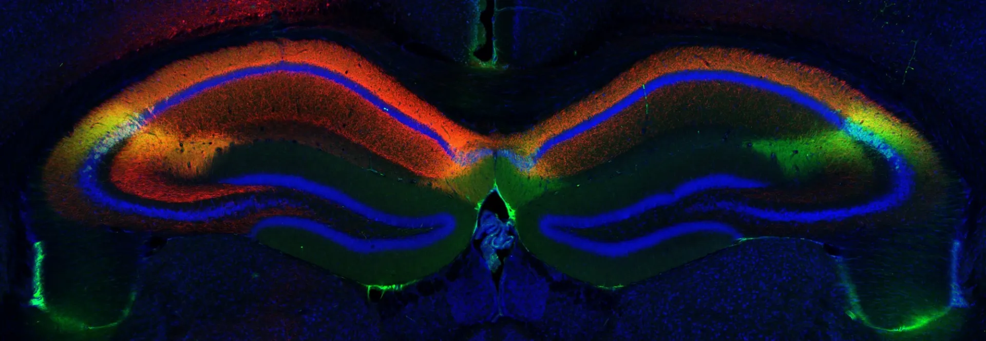
coronal section of the brain with both CA2 regions labeled (RGS14 staining, in green) and axonal projections of one CA2 region (in red, marking virally-expressed GFP)